Whole-heart electrophysiology simulation
Heart Activation Map
An interactive 3D view of how each heartbeat's electrical impulse spreads from the sinus node through the atria, AV node, His–Purkinje system and ventricular walls, with the 12-lead ECG that the beat produces drawn beneath it on the same time axis.
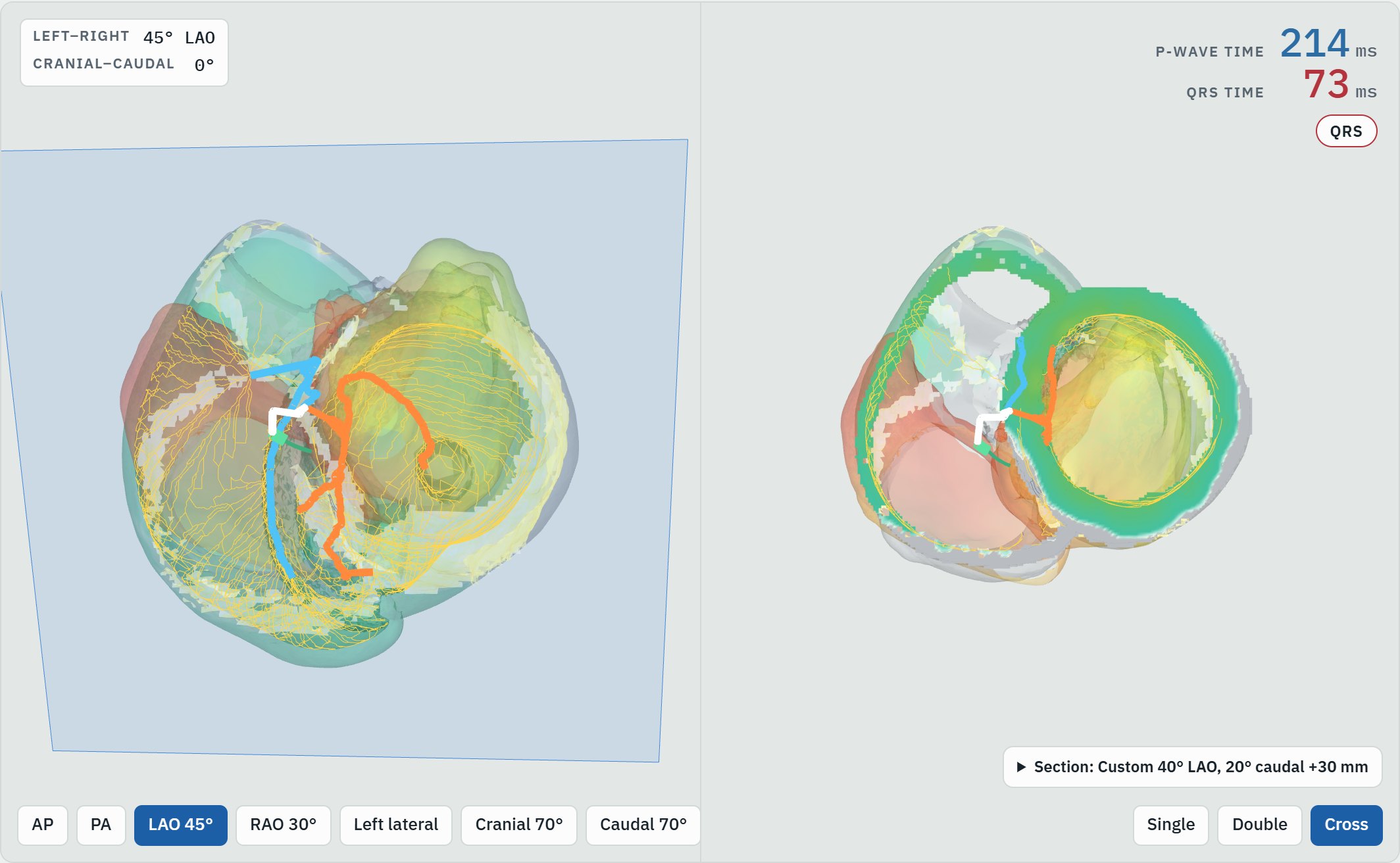
Conduction
- Normal conduction
- Left and right bundle branch block
- Premature ventricular contractions from the RVOT, LVOT, mid LV septum and mid LV lateral wall
Pacing
- Dual chamber: RV apical septum, deep septal, or left bundle branch area pacing (LBBAP)
- Cardiac resynchronization: biventricular, LBBAP-CRT and LOT-CRT
- CRT ECG measures, including QRS duration and QRS area
Hearts and views
- A normal heart and a dilated cardiomyopathy heart
- Single, double (90° apart) and cross-section views
- Fluoroscopy-style angle readout and preset views
- Movie recording of the atrial, ventricular or full sequence
Using the viewer
- Choose a heart and a conduction or pacing scenario from the panel on the right.
- Press Play, or drag the time slider, to watch the wavefront move. Gray tissue is still at rest, and the white band is the moving front.
- Drag to rotate and scroll to zoom. The Cross layout cuts through the walls on a sagittal, coronal, transverse or custom plane.
- The ECG strip below the heart follows the same time cursor; choose the lead to display.
About the model
The heart sits inside a human torso model built from medical imaging, so the ECG reflects where the heart lies in the chest. Activation and ECGs are computed offline by a cardiac electrophysiology simulation and shown here as precomputed results; the viewer replays them in your browser.
For research and education. The viewer is not a medical device and is not intended for diagnosis or treatment decisions.
Slides for discussion · October 20, 2026
Cardiac Pacing 2026: Indications, Lead Strategy, and Outcomes
Current guidelines and trial evidence in 69 slides: reading conduction disease on the ECG, pacing for bradycardia, resynchronization and left bundle branch area pacing, pacing around valve procedures and surgery, what each pacing site does to LVEF, and leadless pacing.